


Stomach cancer in dogs is relatively rare, although it does happen from time to time. Stomach tumors are usually malignant (cancerous) and can spread to other organs. Most stomach tumors are not cancerous, but even benign tumors can cause problems if they grow large or cause an obstruction in the digestive tract.
What is a Stomach Tumor?
The cells that make up the stomach proliferate abnormally and replicate in an uncontrolled manner, resulting in stomach cancer in dogs. Stomach tumors mainly develop from cells in the stomach’s inner lining or the muscle that surrounds it.
Stomach tumors can be benign (non-cancerous) or malignant (cancerous). Malignant tumors are invasive and can spread (spread to other areas of the body). The majority of stomach tumors are cancerous.
Stomach cancer in dogs are rare. The most prevalent cancer in dogs is leiomyosarcoma, whereas the most common cancer in cats is lymphoma. Lymphoma, smooth muscle tumors (leiomyomas and leiomyosarcomas), adenocarcinomas, mast cell tumors, fibrosarcomas, plasmacytomas, and gastrointestinal stromal tumors (GISTs) are all examples of stomach tumors in dogs. Dogs can develop benign adenomatous polyps on occasion.
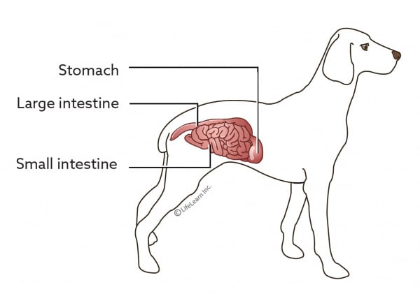
Gastric (stomach) tumors are rare in dogs.
Cause of Stomach Cancer
It’s difficult to say why a particular dog might get this, or any other tumor or disease. Only a small percentage of tumors and malignancies have a single identified etiology. The majority appear to be caused by a complicated combination of risk factors, some of which are environmental in nature and others which are genetic or hereditary. Age, sex, and breed all appear to be risk factors for stomach cancers.
Stomach tumors tend to develop in older dogs. Males are generally at higher risk than females, both for benign and malignant tumors. Certain breeds are at an increased risk, including Beagles for leiomyoma, and Chow Chows, Rough Collies, Staffordshire Terriers, Belgian Shepherds, Norwegian Lundehunds, and Dutch Tervueren Shepherds for adenocarcinoma. This may indicate a genetic predisposition.


In dogs, gastric adenocarcinoma has been linked to the long-term feeding of nitrosamines (a type of chemical found in many foods) in the diet.
Signs of Stomach Tumors
Stomach tumor symptoms appear gradually over weeks to months. Chronic intermittent vomiting, a lack of appetite, lethargy, and weight loss are among them. It’s possible that the vomit will be bloody or have a “coffee grounds” appearance.
This is due to bleeding caused by tumor ulceration (opening). The stool may become blackish as a result of the bleeding. Anemia (few circulating red blood cells) can cause pale gums as a result of chronic bleeding. Excessive salivation is a symptom of nausea that can be experienced on occasion.
Gastric leiomyomas or leiomyosarcomas (muscle tumors) in dogs can cause blood glucose levels to drop below normal (hypoglycemia). This is a form of paraneoplastic syndrome, which occurs when cancer cells release compounds that interfere with the operation of other organs.
Restlessness, weakness, trembling, confusion, and convulsions are all symptoms of low blood glucose (blood sugar). Tumor-related nephrogenic diabetes insipidus is another paraneoplastic condition associated with leiomyomas and leiomyosarcomas. Excessive drinking and urination are symptoms of this illness.
Dogs with adenomatous polyps often have no symptoms of cancer.
Diagnosing Stomach Cancer in Dogs
In elderly dogs or cats with a history of persistent vomiting, lack of appetite, and weight loss, your veterinarian may suspect stomach cancer. A physical examination yields a variety of results. Your veterinarian may be able to palpate (feel) a gastric mass or thickening if your pet’s stomach is noticeably enlarged (sometimes painful). An examination of the abdomen may indicate swollen lymph nodes.
Bloodwork and urinalysis are helpful to find the changes associated with the paraneoplastic syndromes.”
Blood tests, urinalysis, imaging, endoscopy, or surgery and a biopsy will be performed by your veterinarian. Blood tests and urinalysis can be used to detect alterations linked to paraneoplastic disorders. X-rays may reveal a thicker gastric wall or stomach displacement, while specialist imaging (a barium swallow) may reveal ulcers, restricted stomach movement, or gastric obstruction.
Ultrasound can be useful for examining the layers of the stomach wall and obtaining an ultrasound-guided fine needle or core needle biopsy. This process entails suctioning a sample of cells directly from the tumor using a tiny needle and syringe and depositing them on a slide. The slide is subsequently examined under a microscope by a veterinary pathologist.
Endoscopy, a procedure that uses an endoscope (a thin tube with a light and tiny camera at the end through which forceps can be passed to take tissue samples) to diagnose the presence of a tumor and collect biopsy samples, can be helpful in determining the presence of a tumor and collecting biopsy samples.
These samples, like needle biopsies, are not always useful for diagnosis, and a surgical biopsy may be required to achieve a definite (correct) diagnosis. Surgical biopsies can be obtained using laparoscopy, a technique involving the use of a laparoscope (a narrow, tube-like tool with a light and lens), or laparotomy, a procedure involving the use of a laparoscope (a small, tube-like instrument with a light and lens) (a surgery to open the abdomen).
A veterinary pathologist examines the biopsies under a microscope to determine the type of cancer. This is referred to as histology. Histopathology can not only help with diagnosis, but it can also predict how the tumor will behave in the future.
How Does this Cancer Typically Progress?
The type of tumor and how it affects the body determine how stomach cancer grows. Tumors grow at different rates. Some grow slowly, while others grow quickly. Both benign and malignant tumors will continue to grow if they are not treated, interfering with stomach function and increasing the risk of ulceration or gastric obstruction. Ulcerative tumors can cause perforation of the stomach, allowing stomach contents to pour into the abdomen, resulting in a life-threatening infection (septic peritonitis).
Because most stomach tumors are cancerous, they spread to other parts of the body, including the lymph nodes, liver, and lungs, as well as other organs including the abdomen’s inner lining. Staging (looking for potential spread to other parts of the body) is extremely advised when there’s a probability of metastasis.
Blood tests, urinalysis, X-rays of the lungs, and potentially an abdomen ultrasound are all possible tests. More advanced imaging techniques, such as computed tomography (CT) scans or magnetic resonance imaging (MRI), are occasionally utilized (MRI). If any lymph nodes are larger or feel abnormal, further sampling may be done to see if there is any spread. The most appropriate plan of care is set in motion through staging.
What are the Treatments for This Type of Tumor?
The type of stomach tumor and the extent to which it has grown and spread determine the treatment options. Surgery is the preferred treatment for most stomach tumors. The surgical excision of metastasized tumors is primarily for palliative purposes, to alleviate symptoms and improve quality of life.
In many cases, the long-term outlook is usually bleak, with surgery offering just a few months of relief until the metastatic growths become problematic or the tumor grows back. Chemotherapy may be used in conjunction with surgery in some circumstances.
Gastric lymphoma in dogs and cats can be localized (growing as a mass in one location) or diffuse (spread out as a general thickening of the stomach wall). Surgery is advised if it is possible, as it will help alleviate the signs of cancer, but typically lymphoma is treated with chemotherapy whether or not the tumor is removed.
Sometimes chemotherapy is the preferred treatment approach with gastric lymphoma in dogs and cats. In some cases, radiation therapy may also be recommended.
In the case of muscle tumors, when the tumor is removed, the signs of paraneoplastic syndrome will resolve. After surgery, smaller, more frequent meals may be necessary.
Final Thoughts
Depending on the type of tumor, whether it has migrated to other parts of the body, the number of tumors present, and whether all cancer can be removed, the prognosis can range from excellent to bad. The degree of debilitation (e.g., weight loss and malnourishment) or other health concerns your pet has can sometimes restrict the prognosis. Your veterinarian or veterinary oncologist can advise you on the best course of action for your pet.
2 thoughts on “Alarming Signs of Stomach Cancer in Dogs”