
Tissue growth and replication abnormalities in the esophagus, the muscular tube that connects the throat to the stomach, are characterized by abnormal cell proliferation and dysregulation of cell replication. In most cases, esophageal tumors develop from cells in the inner lining of the esophagus or from muscle that surrounds the inner lining.
Esophageal tumors are classified as either benign (noncancerous) or malignant (cancerous). Malignant tumors are aggressive and prone to spreading throughout the body (spread to other areas of the body). The vast majority of esophageal tumors are malignant.
Esophageal tumors are extremely rare in dogs and cats. The most common types of esophageal tumors include leiomyosarcomas (dogs), fibrosarcomas, osteosarcomas, and undifferentiated sarcomas, all of which are malignant. Benign tumors can also occur and include leiomyomas (the most common) and plasmacytomas. Esophageal tumors are mostly found in the lower esophagus in dogs.
Cause of Esophageal Cancer in Dogs
It is difficult to determine why a certain dog may get this tumor or cancer, or any other type of cancer for that matter. Only a small percentage of tumors and malignancies have a single identifiable etiology. Most appear to be caused by a complicated combination of risk factors, some of which are environmental in nature and others which are genetic or hereditary in nature.
The only known cause of esophageal fibrosarcoma, osteosarcoma, and undifferentiated sarcoma in dogs are spirocercosis and adenovirus infection. Spirocercosis is an infection caused by the roundworm Spirocerca lupi, which can be found in tropical and subtropical regions of the world.
As each S. lupi worm settles into the esophagus’s wall, it causes an inflammatory nodule to grow around it, causing the worm to spread. Sarcomas are inflammatory nodules that can progress to malignant transformation and eventually become cancerous tumors. There are no recognized causes of esophageal tumors, with the exception of spirocercosis, at this time.
What are the Signs of Esophageal Tumors?
When an esophageal tumor is present, the indications and symptoms might vary and are directly proportional to its physical size. It becomes increasingly difficult or impossible to transport food from the throat to the stomach as the tumor grows larger and more complex. As a result, the signs and symptoms usually appear gradually and are not recognized until the cancer has progressed significantly.
You may notice that your dog is having increased difficulty or pain swallowing, or that he or she is drooling more frequently. It is possible they will experience coughing or choking while eating. Also possible is the presence of what appears to be vomiting, but which is actually regurgitation (i.e. ejection of the contents of the esophagus, not the stomach).
As your dog’s ability to eat and overall health declines, you may notice that your dog is losing weight or that he or she has stopped eating entirely. Aspiration pneumonia is a serious medical condition that can occur in dogs who have difficulty swallowing, have choking episodes, or regurgitate often. Coughing, fever, and nasal discharge are some of the symptoms of pneumonia.
How is This Cancer Diagnosed?
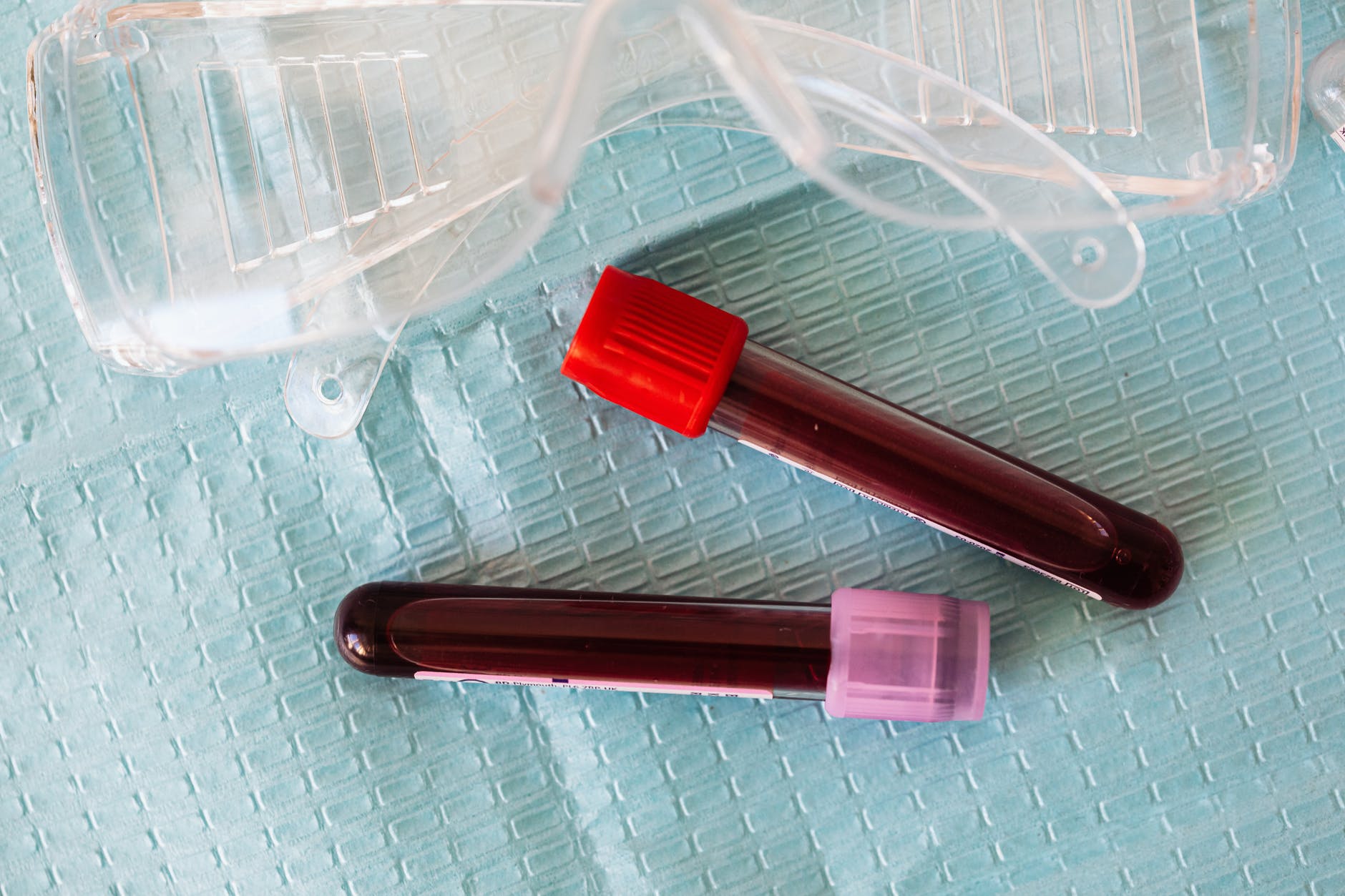
This cancer is typically diagnosed with various forms of imaging and an endoscopic or surgical biopsy.
Chest radiographs (X-rays) may show the presence of a distended esophagus (megaesophagus) and/or an esophageal mass.
If the X-rays shows air, fluid, or food in a distended esophagus, or a mass, your veterinarian may recommend more advanced imaging to see how the esophagus is functioning (e.g., fluoroscopy) or to more clearly find the source of the problem may recommend computed tomography or magnetic resonance imagining.
Chest X-rays may also reveal evidence of metastasis (spread) to the lungs or aspiration pneumonia.
A definitive diagnosis of esophageal cancer may be made via endoscopy and biopsy.
Endoscopy is a procedure that uses an instrument called an endoscope. An endoscope is a thin tube with a light and tiny camera at the end, and through which forceps can be passed to take tissue samples.
The biopsies are then submitted to a pathologist to review under a microscope and determine the type of cancer. This is called histopathology. Histopathology is not only helpful to make a diagnosis but can indicate how the tumor is likely to behave.
Surgical removal of the tumor may be recommended if the endoscopic biopsy is inconclusive. In this case, If the tumor is benign, the surgery could be curative.
Occasionally S. lupi roundworm eggs are detected in the feces of dogs with esophageal sarcomas.
Progression of Esophageal Cancer
Most malignant tumors are locally invasive and metastasize to the nearby lymph nodes or to other areas of the body.
Spirocerca-induced esophageal sarcomas metastasize most frequently to the lungs. Since there is a risk of metastasis, staging (searching for potential spread to other locations in the body) is highly recommended.
This may include:
- Bloodwork
- Urinalysis
- X-rays of the lungs
- Abdominal ultrasound
If any lymph nodes are enlarged or feel abnormal, additional biopsies may be pursued to determine if spread is present.
Another complication of Spirocerca-induced esophageal sarcomas is an unusual condition called hypertrophic osteopathy. Hypertrophic osteopathy is a condition characterized by:
- Painful swelling of the legs
- Lameness
- Lethargy or weakness
It occurs in dogs with certain kinds of infections and cancers.
Treatment of Esophageal Tumors
The treatment of malignant esophageal tumors is difficult because, in most cases, they are diagnosed at an advanced stage, once they have already metastasized (AKA spread to other parts of the body).
Radiation therapy of the upper esophagus (the neck-throat area) may be possible, but not the middle or lower esophagus (the chest area). Radiation generally tends to be of limited value with malignant tumors.
Benign tumors, such as leiomyomas, may be surgically removed via surgery. Prior to becoming cancerous, spirocercosis can be successfully treated with avermectins (a type of drug used to treat parasitic worms).
Understand the Cancer, But Don’t Give Up Hope
Drake Dog Cancer Foundation
Read more:
Esophageal Tumors | VCA Animal Hospital